- HOME
- Enzyme List
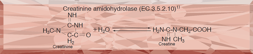
- CNH-211・311 CREATININE AMIDOHYDROLASE
CNH-211・311
CREATININE AMIDOHYDROLASE from Microorganism
PREPARATION and SPECIFICATION
| Appearance | White amorphous powder, lyophilized | |
|---|---|---|
| Activity | GradeⅡ(−211) 450 U/mg-solid or more
GradeⅢ(−311) 150 U/mg-solid or more |
|
| Contaminants | NADH oxidase | ≤5.0×10⁻² % |
| Catalase | ≤2.0 % | |
| Stabilizers | Sucrose, BSA | |
PROPERTIES
| Stability | Stable at −20 ℃ for at least one year (Fig.1,2) |
|---|---|
| Molecular weight | approx. 175,000 1) |
| Isoelectric point | 4.7 3) |
| Michaelis constants | 3.2×10⁻² M (Creatinine), 5.7×10⁻² M (Creatine) |
| Structure | 6 subunits per enzyme molecule (One zinc is bound to each subunit) 3) |
| Inhibitors | Ag+,Hg2+,N-bromosuccinimide, EDTA |
| Optimum pH | 6.5−7.5(Fig.5) |
| Optimum temperature | 70 ℃(Fig.6) |
| pH Stability | pH 7.5−9.0 (5 ℃, 16 hr)(Fig.7) |
| Thermal stability | below 70 ℃ (pH 7.5, 30 min)(Fig.8) |
| Effect of various metals | (Table 1) |
APPLICATIONS
This enzyme is useful for enzymatic determination of creatinine in combination with creatine amidinohydrolase (CRH-211, CRH-221, CRH-229) and sarcosine oxidase (SAO-351) in clinical analysis.
ASSAY
Principle
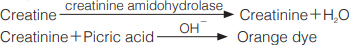
The formation of creatinine-picrate (orange dye based on Jaffe's reaction) is measured at 520 nm by spectrophotometry.
Unit definition
One unit causes the formation of one micromole of orange dye per minute under the conditions detailed below.
Method
Reagents
| A. Creatine solution | 0.1 M [1.49 g creatine (Nacalai tesque)/100 mL of 50 mM phosphate buffer, pH 7.5](Should be prepared fresh) |
|---|---|
| B. NaOH solution | 0.5 N |
| C. Picric acid solution | 1.0 % (W/V) |
| D. Enzyme diluent | 50 mM phosphate buffer, pH 7.5 |
Procedure
1.Pipette 1.0 mL of the substrate solution (A) into a test tube and equilibrate at 37℃ for about 5 minutes.
| Concentration in assay mixture | |
|---|---|
| Phosphate buffer | 45 mM |
| Creatine | 90 mM |
2.Add 0.1 mL of the enzyme solution* and mix.
3.After exactly 10 minutes at 37℃, immediately transfer 0.1 mL(1/11 volume) of the reaction solution to 2.0 mL of NaOH solution (B).
4.Add 1.0 mL of picric acid solution (C) and incubate at 25℃ for 20 minutes.
5.Measure the optical density at 520nm against water (OD test).
At the same time, prepare the blank by transferring 0.1 mL of the reaction solution into NaOH solution (2.0 mL) just after addition of the enzyme solution to ice-cold substrate solution, and carry out the same procedure as the test (procedures 4 and 5; OD blank).
*Dissolve the enzyme preparation in ice-cold 50mM phosphate buffer, at pH 7.5, and dilute to 1.8−2.4U/mL with the same buffer, immediately before the assay.
Calculation
Activity can be calculated by using the following formula :
Volume activity (U/mL) =
-
ΔOD/min (OD test−OD blank)×Vt×11×df
4.65×1.0×t×Vs
= ΔOD×7.33×df
Weight activity (U/mg) = (U/mL)×1/C
| Vt | : Total volume (3.1 mL) |
| Vs | : Sample volume (0.1 mL) |
| 4.65 | : Millimolar extinction coefficient of creatinine-picrate (orange dye) (cm2/micromole) |
| 11 | : Volume of reaction solution (1.1 mL)/Sampling volume (0.1 mL) |
| 1.0 | : Light path length (cm) |
| t | : Reaction time (10 minutes) |
| df | : Dilution factor of enzyme solution |
| C | : Enzyme concentration in dissolution (c mg/mL) |
REFERENCES
1)D.Tsuru; Nucleic Acid and Amino Acids, 35, 31 (1977).
2)D.Tsuru; Rinsho Kensa, 22, 1331 (1978).
3)K.Rikitake, I.Oka, M.Ando, T.Yoshimoto and D.Tsuru; J.Biochem., 86, 1109 (1979).
4)K.Yamamoto, M.Oka, T.Kikuchi and S.Emi; Biosci. Biotech. Biochem., 59 (7), 1331 (1995).
Table 1. Effect of Various Metals on Creatinine amidohydrolase
[The enzyme dissolved in 50mM K-phosphate buffer, pH 7.5 was incubated with each metal (0.2mM) for 30minutes at 25℃.]
-
Metal Residual activity(%) None 100 FeSO4 91.0 FeCl3 93.0 HgCl2 1.4 Zn(OAc)2 79.9 Cu(OAc)2 22.7 Ca(OAc)2 96.8 MnCl2 94.0 -
Metal Residual activity(%) MgCl2 82.8 NiCl2 82.8 CoSO4 78.5 BaCl2 71.2 Pb(OAc)2 88.0 AgNO3 0 CdCl2 80.0
-
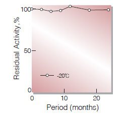
Fig.1. Stability (CNH-211)(Powder form)
(kept under dry conditions)
-
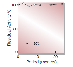
Fig.2. Stability (CNH-311)(Powder form)
(kept under dry conditions)
-
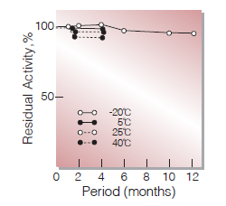
Fig.3. Stability (Powder form)
(kept under dry conditions)
-
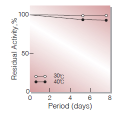
Fig.4. Stability (Liquid form)
(in 50 mM Tris-HCI buffer solution pH7.5)
-
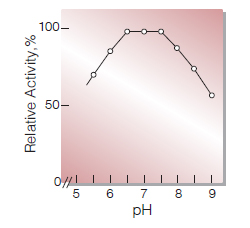
Fig.5. pH-Activity
37℃, 10min-reaction in 50mM buffer solution: pH5.5,acetate;pH6.0-8.0,phosphate;
pH8.5-9.0,carbonate -
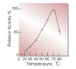
Fig.6. Temperature activity
10min-reaction in 50mM phosphate buffer, pH7.4
-
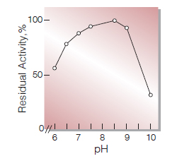
Fig.7. pH-Stability
25℃,16hr-treatment with 50mM buffer solution: pH6.0-8.0,phosphate; pH8.5-9.0, carbonate
-
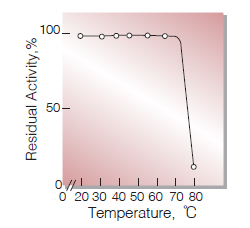
Fig.8. Thermal stability
30min-treatment with 50mM phosphate buffer, pH7.4
活性測定法(Japanese)
1. 原理
生成したクレアチニンをアルカリ性下ピクリン酸と反応させ生じた橙色色素を比色定量する。
2.定義
下記条件下で1分間に1マイクロモルの橙色色素を生成する酵素量を1単位 (U)とする。
3.試薬
- 0.1Mクレアチン溶液〔1.49 gのクレアチン(ナカライテスク製)を50mMリン酸緩衝液 pH7.5に溶解し,100 mLとする〕(用時調製)
- 0.5N NaOH溶液(2.0gのNaOHを蒸留水に溶解し100 mLとする)
- 1.0%ピクリン酸溶液(1.0gのピクリン酸を蒸留水に溶解し100 mLとする)
酵素溶液:酵素標品を予め氷冷し50mMリン酸緩衝液,pH7.5で溶解し,分析直前に同緩衝液で1.8~2.4U/mlに希釈する。
4.手順
1.試験管に基質溶液(A)1.0 mLを採り,37℃で約5分間予備加温する。
2.酵素溶液0.1 mLを加え,反応を開始する。
3.37℃で正確に10分間反応させた後,直ちに反応液0.1 mL(1/11容量)を採り,予め準備したNaOH溶液(B)2.0ml中へ加える。
4.ピクリン酸溶液(C)を1.0 mL加え,25℃で20分間放置後,520nmにおける吸光度を測定する (ODtest)。
5.盲検は氷冷基質溶液に酵素溶液を添加後直ちに反応液の0.1 mLを採り,NaOH溶液(2.0mL)の中へ加えて調製する。以下手順4を操作して吸光度を測定する(ODblank)。
5.計算式
U/mL =
-
ΔOD (OD test−OD blank)×3.1(mL)×11×希釈倍率
4.65×1.0×10(分)×0.1(mL)
| = ΔOD×7.33×希釈倍率 | |
| U/mg | =U/mL×1/C |
| 4.65 | : Creatinine-picrate(Orange dye)のミリモル分子吸光係数(cm2/micromole) |
| 11 | : 反応液容量(1.1mL)/採取液量(0.1mL) |
| 1.0 | : 光路長(cm) |
| C | : 溶解時の酵素濃度(c mg/mL) |
CONTACT
-
For inquiries and cosultations regarding our products, please contact us through this number.
- HEAD OFFICE+81-6-6348-3843
- Inquiry / Opinion