- HOME
- Enzyme List
- COO-311 CHOLESTEROL OXIDASE
COO-311
CHOLESTEROL OXIDASE from Streptomyces sp.

PREPARATION and SPECIFICATION
| Appearance | Yellowish amorphous powder, lyophilized | |
|---|---|---|
| Activity | GradeⅢ 15U/mg-solid or more (contg. approx. 40% of stabilizers) |
|
| Contaminants | Catalase | ≤1.0% |
| Cholesterol esterase | ≤1.0×10⁻²% | |
| Stabilizers | BSA, sugars | |
PROPERTIES
| Stability | Stable at −20℃ for at least one year (Fig.1) |
|---|---|
| Molecular weight | approx. 34,000 (by gel-filtration on Sephadex G-200) |
| Isoelectric point | 5.1±0.1 and 5.4±0.1 |
| Michaelis constant | 4.3×10⁻⁵M (Cholesterol) |
| Inhibitors | Ionic detergents, Hg++, Ag+ |
| Optimum pH | 6.5−7.0(Fig.3) |
| Optimum temperature | 45−50℃(Fig.4) |
| pH Stability | pH 5.0−10.0 (25℃, 20hr)(Fig.5) |
| Thermal stability | below 45℃ (pH 7.5, 15min)(Fig.6) |
| Specificity | (Table 1) |
| Effect of various chemicals | (Table 2) |
APPLICATIONS1〜6)
This enzyme is useful for enzymatic determination of cholesterol in serum when coupled with cholesterol esterase (COE-301, COE-311, COE-313) in clinical analysis.
ASSAY
Principle

The appearance of quinoneimine dye formed when coupled with 4-aminoantipyrine and phenol is measured at 500nm by spectrophotometry.
Unit definition
One unit causes the formation of one micromole of hydrogen peroxide (half a micromole of quinoneimine dye) per minute under the conditions described below.
Method
Reagents
| A. 0.1M K-Phosphate buffer, pH 7.0 | |
|---|---|
| B. Cholesterol solution | To 5.0ml of Triton X-100 on a hot plate or in a water bath, add 500mg of cholesterol and mix with a stirring bar until cholesterol dissolves. Add 90ml of distilled water to the hot cholesterol-Triton X-100 solution by slowly pouring along a stirring bar. Stir and allow to boil for 30 to 60 seconds. The solution will be cloudy. Cool under running water with gentle agitation, the solution will turn clear. Add 4.0g of sodium cholate and dissolve. Fill up the solution to 100ml with distilled water. This solution is stable for about one week at room temperature. If it becomes cloudy, warm slightly while stirring until it clears. |
| C. 4-AA solution | 1.76% (1.76g 4-aminoantipyrine/100ml of H2O) |
| D. Phenol solution | 6.0% (6.0g phenol/100ml of H2O) |
| E. POD solution | Horseradish peroxidase 15,000 purpurogallin units/100ml of buffer (A) |
| F. Enzyme diluent | 20mM K-Phosphate buffer, pH 7.0 contg.0.2% bovine serum albumin |
Procedure
1.Prepare the following working solution (20 tests volume), immediately before use and store on ice in a brownish bottle.
| 51.0ml | Buffer solution | (A) |
|---|---|---|
| 4.0ml | Substrate solution | (B) |
| 1.0ml | 4-AA solution | (C) |
| 2.0ml | POD solution | (E) |
| Concentration in assay mixture | |
|---|---|
| K-Phosphate buffer | 87 mM |
| Cholesterol | 0.89mM |
| 4-Aminoantipyrine | 1.4 mM |
| Phenol | 21 mM |
| Triton X-100 | 0.34 % |
| Sodium cholate | 64 mM |
| BSA | 33μg/ml |
| POD | 5 U/ml |
2.Pipette 2.9ml of working solution into a cuvette (d=1.0cm) and equilibrate at 37℃ for about 3 minutes. Add 0.1ml of Phenol solution (D), mix and keep at 37℃ for another 2 minutes.
3.Add 0.1ml of the enzyme solution* and mix with gentle inversion.
4.Record the increase in optical density at 500nm against water for 3 to 4 minutes in a spectrophotometer thermostated at 37℃, and calculate the ΔOD per minute from the linear portion of the curve (ΔOD test). At the same time, measure the blank rate (ΔOD blank) by using the same method as the test except that the enzyme diluent is added instead of the enzyme solution.
*Dissolve the enzyme preparation in ice-cold enzyme diluent (F), and dilute to 0.1−0.3 U/ml with the same buffer, and store on ice.
Calculation
Activity can be calculated by using the following formula :
Volume activity (U/ml) =
-
ΔOD/min (ΔOD test−ΔOD blank)×Vt×df
13.78×1/2×1.0×Vs
= ΔOD/min×4.499×df
Weight activity (U/mg) = (U/ml)×1/C
| Vt | : Total volume (3.1ml) |
| Vs | : Sample volume (0.1ml) |
| 13.78 | : Millimolar extinction coefficient of quinoneimine dye under the assay conditions (cm2/micromole) |
| 1/2 | : Factor based on the fact that one mole of H2O2 produces half a mole of quinoneimine dye. |
| 1.0 | : Light path length (cm) |
| df | : Dilution factor |
| C | : Enzyme concentration in dissolution (c mg/ml) |
REFERENCES
1)W.Richmond; Clin.Chem., 19, 1350 (1973).
2)H.M.Flegg; Ann.Clin.Biochem., 10, 79 (1973).
3)C.C Alain et al; Clin.Chem., 20, 470 (1974).
4)P.N.Tarbutton and C.R.Gunter; Clin.Chem., 20, 724 (1974).
5)S.Nomoto; Rinsho Kensa, 20, 688 (1976).
6)K.Kameno et al; Jap.J.Clin.Path., 24, 650 (1976).
Table 1. Substrate Specificity of Cholesterol oxidase
-
Substrate(0.1mM) Relative activity(%) Cholesterol 100 Pregnenolone 99.5 β-Cholestanol 75.0 β-Sitosterol 72.2 Stigmasterol 37.2 Dehydroiso-androsterone 24.0 -
Substrate(0.1mM) Relative activity(%) Ergosterol 12.5 Lanosterol ー Testosterone ー Androsterone ー 5β-Pregnone-3α,20α-diol ー
Table 2. Effect of Various Chemicals on Cholesterol oxidase
[The enzyme (1.0U/m) dissolved in 0.1M acetate buffer, pH 6.0 was incubated with each chemical for 1hr at 25℃.]
-
Chemical Concn.(mM) Residual activity(%) None ー 100 Metal salt 2.0 MgCl2 99 CaCl2 98 Ba(OAc)2 100 FeCl3 90 CoCl2 99 MnCl2 99 Zn(OAc)2 99 NiCl2 96 Pb(OAc)2 100 AgNO3 2 HgCl2 27 -
Chemical Concn.(mM) Residual activity(%) PCMB 2.0 100 MIA 2.0 96 NaF 20 92 NaN3 20 92 EDTA 5.0 100 o-Phenanthroline 2.0 92 α,α′-Dipyridyl 2.0 98 Borate 20 100 Triton X-100 0.1% 98 Brij 35 0.1% 99 SDS 0.1% 0 Na-cholate 0.1% 93 Taurocholate 0.1% 95
-
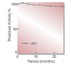
Fig.1. Stability (Powder form)
(kept under dry conditions)
-
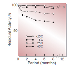
Fig.2. Stability (Powder form)
(kept under dry conditions)
-
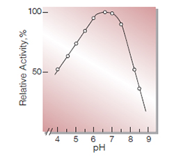
Fig.3. pH-Activity
37℃, in 0.1M buffer solution:pH 4.0-6.0,acetate; pH 5.5-8.5,K-phosphate
-
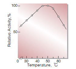
Fig.4. Temperature activity
(in 0.1M K-phosphate buffer, pH7.0)
-
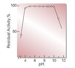
Fig.5. pH-Stability
25℃, 20hr-treatment with 50mM buffer solution:pH4.0-6.0, acetate;pH6.0-8.5, K-phosphate; pH8.5-11.0,K2CO3-NaHCO3
-
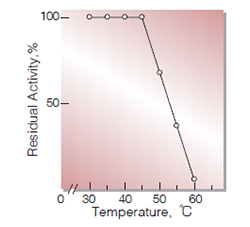
Fig.6. Thermal stability
15min-treatment with 50mM Kphosphate buffer,pH7.0
活性測定法(Japanese)
1. 原理
4-Aminoantipyrineとフェノールの酸化縮合生成物であるQuinoneimine色素を500nmで測定し,上記反応で生成したH2O2量を定量する。
2.定義
下記条件下で1分間に1マイクロモルのH2O2を生成する酵素量を1単位(U)とする。
3.試薬
- 0.1M K-リン酸緩衝液,pH7.0
- コレステロール溶液〔5.0mlのTriton X-100に500mgのコレステロールを添加し,ヒーター上で攪拌溶解する。これに90mlの蒸留水を静かに添加し,攪拌混合後,ヒーター上で30~60秒煮沸する(溶液は濁る)。次いでゆるやかに攪拌しながら流水中で冷却し(溶液は清澄化する,これに4.0gのコール酸ナトリウム塩(ナカライテスク製)を添加して攪拌溶解させた後蒸留水で最終液量を100㎖とする〕(溶液は室温で少なくとも1週間は保存可能,もし保存中に濁る場合は,攪拌しながら加温清澄化すれば良い)
- 4-AA水溶液:1.76%(4-アミノアンチピリン1.76gを水に溶解して100mlとする)
- フェノール水溶液:6.0% (フェノール6.0gを水に溶解して100mlとする)
- POD溶液:Peroxidase 150mg(100プルプロガリン単位/mg)を100mlの緩衝液(A)に溶解する。
酵素溶液:酵素標品を予め氷冷した0.2%のBSAを含む20mM K-リン酸緩衝液, pH7.0で溶解し,同緩衝液で0.1~0.3 U/mlに希釈する。
4.手順
1.下記反応混液を調製する (褐色瓶にて氷冷保存)。
| 51.0ml | K-リン酸緩衝液 | (A) |
| 4.0ml | 基質溶液 | (B) |
| 1.0ml: | 4-AA水溶液 | (C) |
| 2.0ml | POD溶液 | (E) |
2.反応混液2.9mlをキュベット(d=1.0cm)にとり,37℃で約3分間予備加温し,0.1mlのフェノール水溶液を加えて更に2分間加温する。
3.酵素溶液0.1mlを加え,ゆるやかに混和し,水を対照に37℃に制御された分光光度計で500nmの吸光度の増加を3〜4分間記録し,その直線部分から1分間あたりの吸光度変化を求める(ΔOD test)。
4.盲検は反応混液に,酵素溶液の代りに酵素希釈液(0.2% BSAを含む20mM K-リン酸緩衝液,pH7.0)を0.1ml加え,上記同様に操作を行って1分間当りの吸光度変化を求める(ΔODblank)。
5.計算式
U/ml =
-
ΔOD/min (ΔOD test−ΔOD blank)×3.1(ml)×希釈倍率
13.78×1/2×1.0×0.1(ml)
| = ΔOD/min×4.499×希釈倍率 | |
| U/mg | =U/ml×1/C |
| 13.78 | : Quinoneimine色素の上記測定条件下でのミリモル分子吸光係数 (cm2/micromole) |
| 1/2 | : 酸素反応で生成したH2O2の2分子のから形成するQuinoneimine色素は1分子である事による係数。 |
| 1.0 | : 光路長(cm) |
| C | : 溶解時の酵素濃度(c mg/ml) |
CONTACT
-
For inquiries and cosultations regarding our products, please contact us through this number.
- HEAD OFFICE+81-6-6348-3843
- Inquiry / Opinion