- HOME
- Enzyme List
- G3O-321 L-α-GLYCEROPHOSPHATE OXIDASE
-
G3O-321
L-α-GLYCEROPHOSPHATE OXIDASE from Microorganism
PREPARATION and SPECIFICATION
| Appearance | Yellowish amorphous powder, lyophilized | |
|---|---|---|
| Activity | GradeⅢ 15 U/mg-solid or more | |
| Contaminants | Lactate oxidase | ≤ 2.0×10⁻⁴ % |
| Adenosine triphosphatase | ≤ 2.0×10⁻⁴ % | |
| Stabilizers | Amino acids , FAD | |
PROPERTIES
| Stability | Stable at −20 ℃ for at least one year(Fig.1) |
|---|---|
| Molecular weight | approx. 67,000 (by SDS-PAGE) |
| Isoelectric point | 4.6±0.1 |
| Michaelis constant | 1.3×10⁻³ M |
| Inhibitors | SH-reagents, ionic detergents, metal ions, etc. |
| Optimum pH | 6.0−7.0(Fig.3) |
| Optimum temperature | 45 ℃(Fig.4) |
| pH Stability | 4.5−8.5 (25 ℃, 20 hr)(Fig.5) |
| Thermal stability | below 45 ℃ (pH 6.5,15 min)(Fig.6) |
| Effect of various chemicals | (Table 1) |
APPLICATIONS
This enzyme is useful for enzymatic determination of triglyceride in combination with lipoprotein lipase (LPL-311, LPL-314) and glycerokinase (GYK-301, GYK-311) in clinical analysis.
ASSAY
Principle


The formation of quinoneimine dye is measured at 555 nm by spectrophotometry.
Unit definition
One unit causes the formation of one micromole of hydrogen peroxide (half a micromole of quinoneimine dye) per minute under the conditions detailed below.
Method
Reagents
| A. D, L-α-Glycerophosphate solution | 1.5 M: Weigh out 48.63 g of D,L-α-glycerophosphate (disodium salt; MW = 324.17), dissolve in 60 mL of H2O. and, after adjusting the pH to 6.5 ± 0.05 at 25 ℃ with 4.0 N HCl, make up to 100 mL with H2O (Stable for 2 weeks if stored at 0−4 ℃). | |
|---|---|---|
| B. PIPES-NaOH buffer, pH 6.5 | 0.5 M: Weigh out 15.12 g of PIPES (MW = 302.36), suspend in 60 mL of H2O, dissolve in 10 N NaOH, and, after adjusting the pH to 6.5 ± 0.05 at 25 ℃ with 10 N NaOH, make up to 100 mL with H2O (stable for 2 weeks if stored at 0−4 ℃). | |
| C. 4-AA solution | 28 mM: 569 mg of 4-aminoantipyrine (MW = 203.25) / 100 mL of H2O (stable for 1 week if stored at 4 ℃ in a brownish bottle). | |
| D. EHSPT(TOOS) solution | 20 mM: 591 mg of N-ethyl-N-(2-hydroxy-3-sulfopropyl)-m-toluidine (MW = 295.3) / 100 mL of H2O (stable for 1 week if stored at 4℃ in a brownish bottle). | |
| E. Peroxidase solution | 0.05 %: 50 mg of peroxidase (110 purpurogallin units / mg) / 100 mL of H2O (should be freshly prepared). | |
| F. Enzyme diluent | 20 mM PIPES-NaOH buffer, pH 6.5 containing 0.5 M NaCl | |
Procedure
1. Prepare the following working solution (for 40 tests) in a brownish bottle and store on ice).
| 40 mL | Substrate solution | (A) |
| 40 mL | PIPES-NaOH buffer, pH 6.5 | (B) |
| 5 mL | 4-AA solution | (C) |
| 5 mL | EHSPT solution | (D) |
| 10 mL | Peroxidase solution | (E) |
| Concentration in assay mixture | |
|---|---|
| PIPES-NaOH buffer | 193 mM |
| NaCl | 19.2 mM |
| D,L-α-Glycerophosphate | 577 mM |
| 4-Aminoantipyrine | 1.3 mM |
| EHSPT | 0.96 mM |
| Peroxidase | ca.5.3 U/mL |
2. Pipette 2.5 mL of working solution into a cuvette (d = 1.0 cm) and equilibrate at 30 ℃ for about 5 minutes.
3. Add 0.1 mL of the enzyme solution* and mix by gentle inversion.
4. Record the increase in optical density at 555 nm against water for 3 to 4 minutes with a spectrophotometer thermostated at 30 ℃, and calculate the ΔOD per minute from the initial linear portion of the curve (ΔOD test).
At the same time, measure the blank rate (ΔOD blank) using the same method as the ΔOD test except that the enzyme diluent is added instead of enzyme solution.
*Dissolve the enzyme preparation in ice-cold enzyme diluent (F), dilute to 0.05−0.2 U/mL with the same buffer and store on ice.
Calculation
Activity can be calculated by using the following formula :
Volume activity (U/mL) =
-
ΔOD/min (ΔOD test−ΔOD blank)×Vt×df
29.9×1/2×1.0×Vs
= ΔOD/min×1.739×df
Weight activity (U/mg) = (U/mL)×1/C
| Vt | : Total volume (2.6 mL) |
| Vs | : Sample volume (0.1 mL) |
| 29.9 | : Millimolar extinction coefficient of quinoneimine dye under the assay condition (cm2/micromole) |
| 1/2 | : Factor based on the fact that one mole of H2O2 produces half a mole of quinoneimine dye |
| 1.0 | : Light path length (cm) |
| df | : dilution factor |
| C | : Enzyme concentration in dissolution (c mg/mL) |
Table 1. Effect of Various Chemicals on L-α-Glycerophosphate oxidase
[The enzyme dissolved in 0.1 M PIPES-NaOH buffer, pH 7.0 (10 U/mL) was incubated at 25 ℃ for 1 hr.]
-
Chemical Concn.(mM) Residual
activity(%)None - 100 Metal salt Metal salt 2.0 MgCl2 100.0 CaCl2 96.5 BaCl2 99.5 FeSO4 67.4 FeCl3 73.1 CoCl2 99.5 MnCl2 100.1 ZnSO4 96.4 NiCl2 97.9 AgNO3 93.1 CuSO4 84.7 MIA 2.0 98.4 NaF 2.0 99.4 -
Chemical Concn.(mM) Residual
activity(%)NaN3 20 100.2 EDTA 50 99.6 o-Phenanthroline 2.0 101.6 α,α′-Dipyridyl 2.0 100.0 Borate 20 101.8 IAA 2.0 98.7 NEM 2.0 99.8 Hydroxylamine 2.0 100 Triton X-100 0.10 % 110.6 Brij 35 0.10 % 108.9 Tween 20 0.10 % 99.1 Span 20 0.10 % 103.7 Na-cholate 0.10 % 105.8 SDS 0.05 % 3.0 DAC 0.05 % 71.8
MIA, Monoiodoacetate; EDTA, Ethylenediaminetetraacetate; IAA, Iodoacetamido;
NEM, N-Ethylmaleimide; SDS, Sodium dodecyl sulfate; DAC, Dimethylbenzylalkylammonium chloride.
-
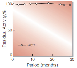
Fig.1. Stability (Powder form)
(Kept under dry form)
-
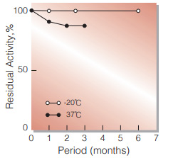
Fig.2. Stability (Powder form)
(Kept under dry form)
-
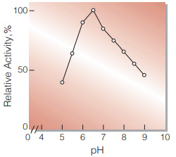
Fig.3. pH-Activity
0.1 M buffer solution:pH 5.0-6.5 ,MES; pH 6.5-7.5,PIPES-NaOH;7.5-9.0,Tris-HCl
-
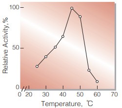
Fig.4. Temperature activity
(in 0.2 M PIPES-NaOH buffer,pH 6.5)
-
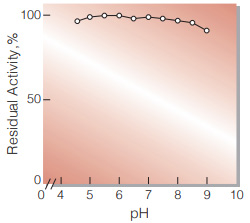
Fig.5. pH-Stability
25 ℃,20 hr-treatment with 0.1 M buffer solution; pH 4.5-6.0,acetate;pH 6.0-8.0,K-phosphate; pH 8.0-9.0,Tris-HClenzyme concn.:20 U/mL,
-
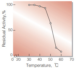
Fig.6. Thermal stability
15 min-treatment with 0.1 M PIPES-NaOH buffer,pH 6.5, enzyme concn.:20 U/mL
活性測定法(Japanese)
1. 原理
4-AminoantipyrineとEHSPTの酸化縮合生成物であるQuinoneimine色素を555nmで測定し, 上記反応で生成したH2O2量を定量する。
2.定義
下記条件下で1分間に1マイクロモルのH2O2を生成する酵素量を1単位(U)とする。
3.試薬
- 1.5Mグリセロリン酸溶液[48.63gのD,L-αグリセロリン酸・2Na(MW=324.17)を約60 mLの蒸留水で溶解後, 4.0N HClでpHを6.5±0.05に調整(25℃)し, 蒸留水で100 mLとする。](0〜4℃保存で2週間は使用可能)
- 0.5M PIPES-NaOH緩衝液,pH6.5[15.12gのPIPES(MW=302.36)を約60 mLの蒸留水で溶解しながら, 10N NaOHでpHを6.5±0.05に調整(25℃)し, 蒸留水で100 mLとする。](0〜4℃保存で2週間は使用可能)
- 28mM 4-AA水溶液[569mgの4-アミノアンチピリン(MW=203.25)を100 mLの蒸留水に溶解する。](褐色瓶中4℃保存で1週間は使用可能)
- 20mM EHSPT(TOOS)水溶液[591mgのEHSPT(MW=295.3)を100 mLの蒸留水に溶解する。(褐色瓶中4℃保存で1週間は使用可能)
- POD溶液[50mgのペルオキシダーゼ(POD)(110プルプロガリン単位/mg)を100 mLの蒸留水に溶解する。](褐色瓶中で4℃保存)(用時調製)
酵素溶液:酵素標品を予め氷冷した0.5M NaCl含む20mM PIPES-NaOH緩衝液,pH6.5で溶解(約1mg/mL)し, 分析直前に同緩衝液で0.05〜0.20U/mLに希釈する。
4.手順
1.下記反応混液を調製する。(褐色瓶にて氷冷保存)
| 40 mL | 基質溶液 | (A) |
| 40 mL | 緩衝液 | (B) |
| 5 mL | 4-AA水溶液 | (C) |
| 5 mL | EHSPT水溶液 | (D) |
| 10 mL | POD水溶液 | (E) |
2.反応混液2.5 mLをキュベット(d=1.0cm)にとり,30℃で約5分間予備加温する。
3.酵素溶液0.1 mLを添加し, ゆるやかに混和後, 水を対照に30℃に制御された分光光度計で555nmの吸光度変化を3〜4分間記録し, その初期直線部分から1分間当りの吸光度変化を求める(ΔODtest)。
4.盲検は反応混液①に酵素溶液の代りに酵素希釈液(0.5M NaCl含む20mM PIPES-NaOH緩衝液, pH6.5)0.10 mLを加え, 上記同様に操作を行い,1分間当りの吸光度変化を求める(ΔODblank)。
5.計算式
U/mL =
-
ΔOD/min (ΔOD test−ΔOD blank)×2.60(mL)×希釈倍率
29.9×1/2×1.0×0.1
| = ΔOD/min×1.739×希釈倍率 | |
| U/mg | = U/mL×1/C |
| 29.9 | : Quinoneimine色素の上記測定条件下でのミリモル分子吸光係数(cm2/micromole) |
| 1/2 | : 酵素反応で生成したH2O21分子から形成するQuinoneimine色素は1/2分子である事による係数 |
| 1.0 | : 光路長(cm) |
| C | : 溶解時の酵素濃度(c mg/mL) |
CONTACT
-
For inquiries and cosultations regarding our products, please contact us through this number.
- HEAD OFFICE+81-6-6348-3843
- Inquiry / Opinion