- HOME
- Enzyme List
- LCD-211 D-LACTATE DEHYDROGENASE
-
LCD-211
D-LACTATE DEHYDROGENASE from Microorganism

PREPARATION and SPECIFICATION
| Appearance | White amorphous powder, lyophilized | |
|---|---|---|
| Activity | GradeⅡ 400 U/mg-solid or more | |
| Contaminants | NADH oxidase | ≤1.0×10⁻³ % |
| Malate dehydrogenase | ≤1.0×10⁻² % | |
| GOT | ≤5.0×10⁻³ % | |
| GPT | ≤5.0×10⁻³ % | |
| Myokinase | ≤1.0×10⁻² % | |
| Pyruvate kinase | ≤1.0×10⁻³ % | |
PROPERTIES
| Stability | Stable at −20 ℃ for at least one year(Fig.1) |
|---|---|
| Molecular weight | approx. 140,000 (by gel filtration) |
| Isoelectric point | 4.0 |
| Michaelis constant | 1.6×10⁻⁴ M (pyruvate, pH 7.0) |
| Inhibitors | Ag+, Hg2+, SH-reagents |
| Optimum pH | 6.0−7.0(Fig.3) |
| Optimum temperature | 35−40 ℃(Fig.4) |
| pH Stability | pH 5.0−9.0 (25 ℃, 48 hr)(Fig.5) |
| Thermal stability | below 45 ℃ (pH 7.0, 15 min)(Fig.6) |
| Effect of various chemicals | (Table 1) |
APPLICATIONS
This enzyme is useful for enzymatic determination of numerous metabolites, such as ATP, ADP, glucose, creatinine, pyruvate, lactate and glycerol, and the activities of enzymes such as GPT, PK and CPK, in combination with related enzymes.
ASSAY
Principle

The formation of NADH is measured at 340 nm by spectrophotometry.
Unit definition
One unit causes the oxidation of one micromole of NADH per minute under the conditions detailed below.
Method
Reagents
| A. Pyruvate solution | 5.0 mM[5.50 mg sodium pyruvate (MW=110)/10 mL of H2O](Should be prepared fresh) | |
|---|---|---|
| B. K-Phosphate buffer, pH 7.4 | 1.0 M | |
| C. NADH solution | 1.0 mM[7.63 mg NADH・2Na (MW=763)/10 mL of H2O](Should be prepared fresh) | |
| D. Enzyme diluent | 0.1 M K-phosphate buffer, pH 7.4 contg. 0.1 % of BSA | |
Procedure
1. Prepare the following working solution for 10 tests in a brownish bottle, immediately before use and store on ice.
| 3.0 mL | Substrate solution | (A) |
| 2.0 mL | Potassium phosphate buffer, pH 7.4 | (B) |
| 3.0 mL | NADH solution | (C) |
| 22.0 mL | H2O | (D) |
| Concentration in assay mixture | |
|---|---|
| Potassium phosphate buffer | 67 mM |
| Pyruvate | 0.49 mM |
| NADH | 0.098 mM |
| BSA | 16.4 μg/mM |
2. Pipette 3.0 mL of working solution into a cuvette (d = 1.0 cm) and equilibrate at 25 ℃ for approximately 5 minutes.
3. Add 0.05 mL of the enzyme solution* and mix by gentle inversion.
4. Record the decrease in optical density at 340 nm against water for 2 to 3 minutes with a spectrophotometer thermostated at 25 ℃, and calculate the ΔOD per minute from the initial linear portion of the curve (ΔOD test).
At the same time, measure the blank rate (ΔOD blank) using the same method as the test except that the enzyme diluent is added instead of the enzyme solution.
*Dilute the enzyme preparation to 0.2−1.0 U/mL with ice-cold enzyme diluent (D), immediately before the assay.
Calculation
Activity can be calculated by using the following formula :
Volume activity (U/mL) =
-
ΔOD/min (ΔOD test−ΔOD blank)×Vt×df
6.22×1.0×Vs
= ΔOD/min×9.81×df
Weight activity (U/mg) = (U/mL)×1/C
| Vt | : Total volume (3.05 mL) |
| Vs | : Sample volume (0.05 mL) |
| 6.22 | : Millimolar extinction coefficient of NADH (cm2/micromole) |
| 1.0 | : Light path length (cm) |
| df | : Dilution factor |
| C | : Enzyme concentration in dissolution (c mg/mL) |
REFERENCES
1) C.A.Loshon, R.B.McComb, L.W.Bond, G.N.Bowers, Jr.W.H.Coleman and R.H.Gwynn; Clin.Chem., 23, 1576 (1977).
2) H.Taguchi, M.Machida, H.Matsuzawa and T.Ohta; Agric.Biol.Chem., 49 (2), 359 (1985).
3) F.Gasser, M.Doudoroff, and R.Contopoulos; J.Gen.Microbiol. 62, 241 (1970).
Table 1. Effect of Various Chemicals on D-Lactate dehydrogenase
[The enzyme dissolved in 0.1 M K-phosphate buffer, pH 7.4 (20 U/mL) was incubated with each chemical at 25 ℃ for 1 hr.]
-
Chemical Concn.(mM) Residual
activity(%)None - 100 Metal salt 2.0 MgCl2 93.0 CaCl2 99.8 Ba(OAc)2 98.1 FeCl2 87.9 CoCl2 91.5 MnCl2 92.2 ZnSO4 89.6 Cd(OAc)2 91.3 NiCl2 92.6 CuSO4 93.8 Pb(OAc)2 93.2 AgNO3 73.0 HgCl2 0 -
Chemical Concn.(mM) Residual
activity(%)PCMB 2.0 89.3 MIA 2.0 0.1 NaF 2.0 98.3 NaN3 20 94.6 EDTA 5.0 99.2 o-Phenanthroline 2.0 95.3 α,α′-Dipyridyl 1.0 93.7 Borate 50 95.6 IAA 2.0 33.6 NEM 2.0 97.7 Hydroxylamine 2.0 95.6 Triton X-100 0.10 % 121 Brij 35 0.10 % 116 Tween 20 0.10 % 117 Span 20 0.10 % 105 Na-cholate 0.10 % 112 SDS 0.05 % 109 DAC 0.05 % 52.0
Ac, CH3CO; PCMB, p-Chloromercuribenzoate; MIA, Monoiodoacetate; EDTA, Ethylenediaminetetraacetate; IAA, Iodoacetamide; NEM, N-Ethylmaleimide; SDS, Sodium dodecyl sulfate; DAC, Dimethylbenzylalkylammonium chloride.
-
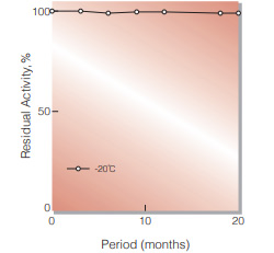
Fig.1. Stability (Powder form)
(kept under dry conditions)
-
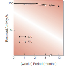
Fig.2. Stability (Powder form)
(kept under dry conditions)
-
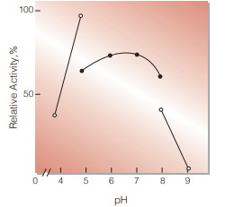
Fig.3. pH-Activity
in 67 mM buffer solution; pH 4-5, acetate;pH 5-8,K-phosphate;pH 8-9 Tris-HCI
-
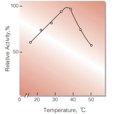
Fig.4. Temperature activity
(in 67 mM K-phosphate buffer, pH 7.4)
-
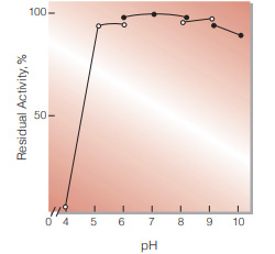
Fig.5. pH-Stability
25 ℃,48hr-treatment with 0.1 M buffer solution: pH 4-6, dimethylgutaric acid-NaOH;pH 6-8, K-phosphate; pH 8-9,Tris-HCI; pH 9-10,glycine-NaOH. enzyme concn.: 10 U/mL
-
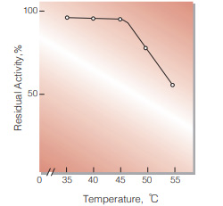
Fig.6. Temperature stability
15 min-treatment with 50 mM K-phosphate buffer,pH 7.0.enzyme concn.: 10 U/mL
活性測定法(Japanese)
1. 原理
NADHの消失量を340nmの吸光度の変化で測定する。
2.定義
下記条件下で1分間に1マイクロモルのNADHが酸化さ れる酵素量を1単位(U)とする。
3.試薬
- 5.0mM ピルビン酸ナトリウム水溶液(用時調製)
- 1.0M K-リン酸緩衝液,pH7.4
- 1.0mM NADH水溶液(用時調製)
酵素溶液:分析直前に酵素標品を予め氷冷した0.1%BSAを含む0.1M K-リン酸緩衝液,pH7.4で溶解(約1mg/ml)し,分析直前に同緩衝液で0.2〜1.0U/mlに希釈する。
4.手順
1.下記反応混液を使用直前に調製する。(褐色瓶にて氷冷保存)
| 3.0ml | 基質溶液 | (A) |
| 2.0ml | K-リン酸緩衝液 | (B) |
| 3.0ml | NADH水溶液 | (C) |
| 22.0ml | H2O |
2.反応混液3.0mlをキュベット(d=1.0cm)に採り,25℃で約5分間予備加温する。
3.酵素溶液0.05mlを添加し,ゆるやかに混和後,水を対照 に25℃に制御された分光光度計で340nmの吸光度 変化を2〜3分間記録し,その初期直線部分から1分間 当りの吸光度変化を求める(ΔOD test)。
4.盲検は反応混液①3.0mlに酵素溶液の代りに酵素希釈液(0.1% BSAを含む0.1M K-リン酸緩衝液, pH7.4)を0.05mlを加え,上記同様に操作を行って,1分間当りの吸光度変化を求める(ΔODblank)。
5.計算式
U/ml =
-
ΔOD/min (ΔOD test−ΔOD blank)×3.05(ml)×希釈倍率
6.22×1.0×0.05(ml)
| = ΔOD/min×9.81×希釈倍率 | |
| U/mg | = U/ml×1/C |
| 6.22 | : NADHのミリモル分子吸光係数(cm2/micromole) |
| 1.0 | : 光路長(cm) |
| C | : 溶解時の酵素濃度(c mg/ml) |
CONTACT
-
For inquiries and cosultations regarding our products, please contact us through this number.
- HEAD OFFICE+81-6-6348-3843
- Inquiry / Opinion