- HOME
- Enzyme List
- XTO-212 XANTHINE OXIDASE
XTO-212
XANTHINE OXIDASE from Microorganism

PREPARATION and SPECIFICATION
| Appearance | Reddish brown amorphous powder, lyophilized | |
|---|---|---|
| Activity | GradeⅡ 10 U/mg-solid or more | |
| Contaminants | Catalase | ≤5 % |
| Adenosine deaminase | ≤1.0×10⁻³ % | |
| Uricase | ≤1.0×10⁻³ % | |
| Purine-nucleoside phosphorylase | ≤5.0×10⁻³ % | |
| Stabilizers | Sodium glutamate, BSA | |
PROPERTIES
| Stability | Stable at −20 ℃ for at least one year (Fig.1) |
|---|---|
| Molecular weight | approx. 160,000 |
| Isoelectric point | 4.0±0.1 |
| Michaelis constant | 4.5×10⁻⁵ M (Xanthine) 7.6×10⁻⁵ M (Hypoxanthine) |
| Inhibitors | Reducing agents, Hg2+, Ag+,MIA |
| Optimum pH | 7.5−8.0 (Fig.3) |
| Optimum temperature | 65 ℃ (Fig.4) |
| pH Stability | pH 6.5−9.0 (25 ℃, 15 hr) (Fig.5) |
| Thermal stability | below 55 ℃ (pH 8.0, 30 min)(Fig.6) |
| Substrate specificity | (Table 1) |
| Effect of various chemicals | (Table 2) |
APPLICATIONS
This enzyme is useful for enzymatic determination of inorganic phosphorus, 5'-nucleotidase and adenosine deaminase, in combination with purine nucleoside phosphorylase (PNP-311) and uricase (UAO-201, UAO-211).
ASSAY
Principle

The formation of uric acid is measured at 293 nm by spectrophotometry.
Unit definition
One unit causes the formation of one micromole of uric acid per minute under the conditions detailed below.
Method
Reagents
| A. Tris-HCl buffer, pH 7.5 | 0.1 M |
|---|---|
| B. Sodium hydroxide solution | 0.025 M |
| C. Xanthine solution | 10 mM[Dissolve 15.2 mg of xanthine (MW=152.11) in 10 mL solution (B)] |
| D. Oxonic acid potassium salt solution | 1 mM (Dissolve 9.75 mg of oxonic acid・K salt in 50 mL H2O) |
| E. Enzyme diluent | 50 mM Tris-HCl buffer, pH 7.5 |
Procedure
1.Prepare the following reaction mixture in a cuvette (d = 1.0cm) and equilibrate at 37 ℃ for approximately 5 minutes.
| 2.24 mL | Tris-HCl buffer, pH 7.5 | (A) |
|---|---|---|
| 0.08 mL | Xanthine solution | (C) |
| 0.08 mL | Oxonic acid solution | (D) |
| Concentration in assay mixture | |
|---|---|
| Tris-HCl buffer | Approx. 89.6 mM |
| Xanthine | 0.32 mM |
| Oxonic acid | 32 μM |
2.Add 0.1 mL of the enzyme solution* and mix by gentle inversion.
3.Record the increase in optical density at 293 nm against water for 3 to 4 minutes with a spectrophotometer thermostated at 37 ℃, and calculate the ΔOD per minute from the initial linear portion of the curve (ΔOD test).
At the same time, measure the blank rate (ΔOD blank) using the same method as in the test except that the enzyme diluent (E) is added instead of the enzyme solution.
*Dissolve the enzyme preparation in ice-cold enzyme diluent (E), dilute to 0.1−0.2 U/mL with the same buffer and store on ice.
Calculation
Activity can be calculated by using the following formula :
Volume activity (U/mL) =
-
ΔOD/min (ΔOD test−ΔOD blank)×Vt×df
12.5×1.0×Vs
= ΔOD/min×2.0×df
Weight activity (U/mg) = (U/mL)×1/C
| Vt | : Total volume (2.5 mL) |
| Vs | : Sample volume (0.1 mL) |
| 12.5 | : Millimolar extinction coefficient of uric acid under the assay condition (㎠/micromole) |
| 1.0 | : Light path length (cm) |
| df | : Dilution factor |
| C | : Enzyme concentration in dissolution (c mg/mL) |
Table 1. Substrate Specificity of Xanthine oxidase (color-system)
-
Substrate(100mM) Relative activity(%) Xanthine 100 Hypoxanthine 18.7 Purine 6.4 Guanine 18.3 Adenosine 0
Table 2. Effect of Various Chemicals on Xanthine oxidase
[The enzyme dissolved in 50 mM Tris-HCl buffer, pH 7.5 (2 U/mL) was incubated with each chemical at 25 ℃ for 2 hr.]
-
Chemical Concn.(mM) Residual
activity(%)None - 100 Metal salt 2.0 MgCl2 78 CaCl2 96 Ba(OAc)2 102 FeCl3 85 CoCl2 100 MnCl2 102 ZnSO4 62 CdCl2 34 NiCl2 91 CuSO4 18 PbCl2 9.7 AgCl 0 HgCl2 1.2 -
Chemical Concn.(mM) Residual
activity(%)PCMB 2.0 24 MIA 2.0 1.1 NaF 2.0 14 NaN3 20 25 EDTA 5.0 95 o-Phenanthroline 2.0 89 α,α′-Dipyridyl 1.0 81 Borate 20 89 IAA 2.0 24 NEM 2.0 91 Triton X-100 0.1 % 99 Brij 35 0.1 % 50 Tween 0.1 % 106 Span 20 0.1 % 110 Na-cholate 0.1 % 115 SDS 0.05 % 111 DAC 0.05 % 78
Ac, CH3CO; PCMB, p-Chloromercuribenzoate; MIA, Monoiodoacetate; EDTA, Ethylenediaminetetraacetate;
IAA, Iodoacetamide; NEM, N-Ethylmaleimide; SDS, Sodium dodecyl sulfate; DAC, Dimethylbenzylalkylammonium chloride.
-
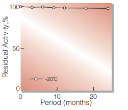
Fig.1. Stability (Powder form)
(kept under dry conditions)
-
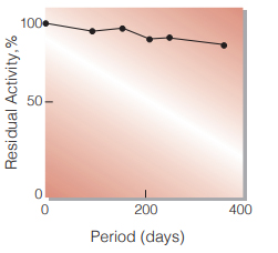
Fig.2. Stability (Liquid form)
in 50 mM buffer solution 4 ℃,pH 6.8 enzyme concn.:60 U/mL
-
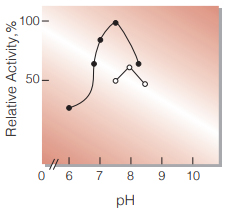
Fig.3. pH-Activity
37 ℃ in 4 min-reaction in 50 mM buffer solution
●:pH 6.0-8.2 K-phosphate
○:pH 7.5-8.5 Tris-HCI -
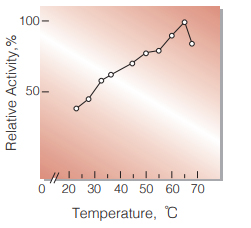
Fig.4. Thermal activity
(in 50 mM Tris-HCl buffer, pH 8.0)
-
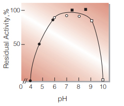
Fig.5. pH-Stability
25 ℃, 15 hr with 50 mM buffer solution
●:pH 4.0-6.0 acetate
○:pH 6.0-8.0 K-phosphate
■:pH 8.0-9.0 Tris-HCI
□:pH 9.0-10.0 Na-K carbonate -
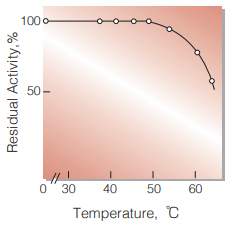
Fig.6. Thermal stability
30 min-treatment with 50 mM Tris-HCI buffer,pH 8.0
活性測定法(Japanese)
1. 原理
尿酸の生成量を293nmにおける吸光度の変化で測定する。
2.定義
下記条件で1分間に1マイクロモルの尿酸を生成する酵素量を1単位(U)とする。
3.試薬
- 0.1M Tris-HCl緩衝液, pH7.5
- 25mM NaOH水溶液
- 10mMキサンチン水溶液〔15.2㎎のキサンチン (MW=152.11)を10mlのNaOHの水溶液(B)で 加温溶解する。〕(4℃保存で2週間は使用可能)
- 1mMオキソン酸・K水溶液〔9.75㎎のオキソン酸・K塩(MW=195.18)を50mlの蒸留水に溶解する。〕(4℃保存で2週間は使用可能)
酵素溶液:酵素標品を予め氷冷した50mM Tris-HCl緩 衝液,pH7.5で溶解し,同緩衝液で0.1- 0.2U/mlに希釈して氷冷保存する。
4.手順
1.下記反応混液をキュベット(d=1.0cm)に調製し,37℃ で約5分間予備加温する。
| 2.24ml | Tris-HCl緩衝液 | (A) |
| 0.08ml | キサンチン水溶液 | (C) |
| 0.08ml | オキソン酸・K水溶液 | (D) |
2.酵素溶液0.1mlを添加し,ゆるやかに混和後,水を対照に37℃に制御された分光光度計で293nmの吸光度変化を求める(ΔODtest)。
3.盲検は反応混液①に酵素溶液の代わりに酵素希釈液(50mM Tris-HCl緩衝液, pH7.5)を0.1ml加え,上記同様に操作を行って,1分間当たりの吸光度変化を求める(ΔODblank)。
5.計算式
U/ml =
-
ΔOD/min (OD test−OD blank)×2.5(ml)×希釈倍率
12.5×1.0×0.1(ml)
| = ΔOD/min×2.0×希釈倍率 | |
| U/mg | = U/ml×1 / C |
| 12.5 | : 尿酸のミリモル分子吸光係数 (cm2/micromole) |
| 1.0 | : 光路長(cm) |
| C | : 溶解時の酵素濃度(c mg/ml) |
CONTACT
-
For inquiries and cosultations regarding our products, please contact us through this number.
- HEAD OFFICE+81-6-6348-3843
- Inquiry / Opinion