


ABOUT US
ACCELERATION OF CREATION
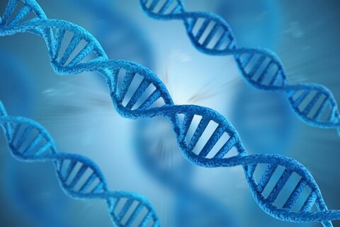
Toyobo is a leading manufacturer of enzymes and reagents for life science research and industrial fields based on our original technologies about PCR enzymes, antibodies, modifying enzymes, magnetic beads and immunoassays.